X-ray

X-ray

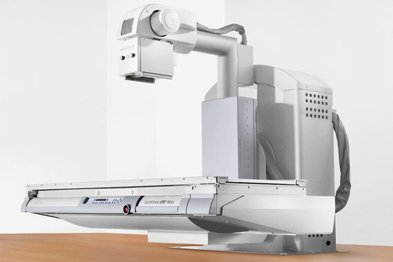
In X-rays, similar to photography with flash light, a short X-ray flash is triggered by applying electricity, which exposes a sensitive film (detector) on the other side of the patient through the patient. The imaged organs cast a shadow on the detector during X-ray, thus important information about lung diseases and bone fractures can be found.

X-ray fluoroscopy

In this examination, similar to video filming, many sequential images are calculated from a series of X-ray flashes by very fast detectors. With the help of contrast medium, information about the gastrointestinal tract can be obtained with this X-ray examination.

Radiology Center - Patientin wird zum Röntgen aufgerufen
A digital X-ray unit that uses a small amount of X-rays to take high-resolution digital images, especially of joints, for diagnosis and treatment planning in orthopedics and trauma surgery. This X-ray unit provides exceptional imaging flexibility in a wide range of settings with patients standing, sitting and lying down.
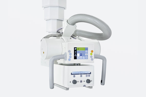
Accurate digital images of internal organs, joints and bones can be made with a small amount of X-ray radiation. If required, short X-ray films (fluoroscopy) can also be taken, for example to examine the mobility of the stomach or intestines.

The different tissues of the body have different densities and thus absorb X-rays to different degrees. Within fractions of a second, an image of the inside of the body is created – with today’s very low radiation exposure (imaging procedure). The Radiology Center’s X-ray equipment provides X-ray images in excellent digital quality immediately after they are taken.
X-rays are also used in computed tomography (CT), but in MRI the images are obtained using magnetic fields and radio waves.

X-rays are produced by the use of high voltage current in X-ray tubes. X-rays are “ionizing” rays, which are rays that can remove electrons from atoms or molecules. X-rays are used only with exact attention to radiation protection and when the examination is clinically justified. The amount of X-rays used varies – depending on the patient and the information required – and is therefore always individually adjusted.
In practical terms, X-rays pass through the human body in much the same way that sunlight passes through paper or flower petals. The different tissues of the human body have different shadowing effects on the X-rays used, so they also appear in different shades of gray, black and white (shading or brightening) on the X-ray image.
X-rays are the radiological method of first choice for all diseases of the musculoskeletal system (e.g. broken bones) or the lungs (e.g. pneumonia).

After registration (with e-card or ID card), you will be seated in one of our waiting areas. From there, the Radiology Center staff will lead you to a changing room. There you will have the opportunity to provide information about your condition, and you will also receive information about the X-ray, computed tomography CT or ultrasound examination.
After the completion of the diagnostic radiology examination, you will receive the images and the information to view the findings online. The billing is done by the elective doctor Dr. Johannes Sailer (elective doctor of all health insurance private).

An X-ray is a procedure in which a short X-ray flash is triggered by an electric current, similar to photography with a flash light, which exposes a sensitive film (detector) on the other side of the patient through the patient. The imaged organs cast a shadow on the detector, and from these shadow images, specialists can deduce the correct information.

The Radiology Center performs the following X-ray examinations:

Fluoroscopy, similar to videography, involves the calculation of many sequential images from a series of X-ray flashes by very fast detectors.

The following examinations are performed at the Radiology Center:

  • Angiography (imaging of blood vessels).
  • Gastric and intestinal examinations

Hip replacements, knee replacements, dental implants, breast implants, heart valves, pacemakers, trauma surgery screws, drills and plates, as well as piercings and tattoos may affect exam results and can only be examined with special equipment settings.

Some patients with certain implants and prostheses must not be allowed in the magnetic resonance imaging room under any circumstances; there would be a danger to the patient’s life.

Please always take your implant ID card with you – if you have one. For your safety, we will clarify whether and how the requested examination, or a better alternative, can be performed.