Sonography

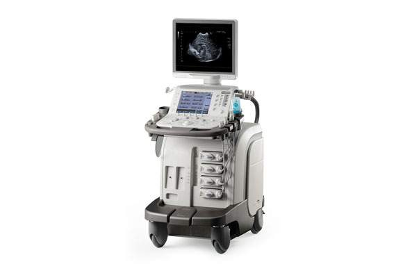
Sonography is the use of ultrasound for imaging. The ultrasound waves are generated and measured with piezoelectric crystals in the transducer. The different tissues of the human body reflect and scatter sound waves (e.g. liver or urinary bladder) to different degrees; images (e.g. abdominal ultrasound) are calculated from the reflected signals. If the ultrasound waves hit a moving surface (such as blood cells in flow), they are reflected with a different frequency (= Doppler effect).

With a so-called Doppler sonography, both flow direction and flow velocity of the blood flow in blood vessels can be determined. The mobility of organs, joints and tendons can also be displayed and assessed in real time with the active assistance of the patient.

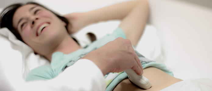
Ultrasound examinations do not use X-rays, so they can be used without hesitation even in pregnant women and children. Of course, you can choose to have your sonography examination performed by Dr. Sailer or Dr. Peloschek (all organs) or Dr. Hoffmann (thyroid, elastography).

  • Sonography thyroid gland
  • Vaginal sonography
  • Sonography uterus
  • Sonography salivary gland
  • Sonography scrotum
  • Sonography shoulder
  • Sonography prostate
  • Sonography kidneys
  • Sonography of the lymph nodes
  • Sonography incl. elastography
  • Sonography inguinal
  • Sonography testicle
  • Sonography hernias
  • Sonography urinary bladder
  • Sonography neck
  • Sonography carotid artery
  • Sonography breast
  • Sonography abdomen
  • Residual urine determination
  • Elastography thyroid gland
  • Doppler sonography pelvic-leg arteries
Only ultrasound waves are used in sonography to generate moving images. With the help of the high-resolution transducer and heavy-wave elastography, pathological changes (inflammations, cancers, thyroid nodules) can be detected and assigned with high accuracy.

Sonography (also echography or colloquially ultrasound sonography) is the use of ultrasound as an imaging procedure for the examination of organic tissue (thyroid, liver, etc.) in medicine and veterinary medicine as well as technical structures. Ultrasound can also be used safely in pregnant women and children for imaging internal organs, and also the gastrointestinal tract (appendicitis, appendicitis). However, air, gas and bone affect the possibilities of ultrasound examination. impenetrable barriers to this procedure.

In sonography, ultrasound waves are generated and measured by piezoelectric crystals in a transducer. The different tissues of the human body reflect and scatter sound waves to different degrees; images are calculated from the reflected signals Sonography Vienna. If the ultrasound waves hit a moving surface (such as blood cells in flow), they are reflected at a different frequency (= Doppler effect). Air, gas and bone form impenetrable barriers to this technique.

With a so-called duplex sonography, both flow direction and flow velocity of the blood stream within a blood vessel can be determined. The mobility of organs, joints and tendons can also be displayed and assessed as an image in real time with the active assistance of the patient.

Ultrasound examinations do not require X-rays, so they can also be used without hesitation in pregnant women and children.

The following ultrasound examinations are performed at the Radiology Center:

  • Abdominal Sonography: Lliver, gallbladder, pancreas, spleen, kidneys, urinary bladder, prostate, uterus, ovaries.
  • Surface Sonography for “small organs” (English: “small parts”): Lymph nodes, thyroid gland, testicles, joints, muscles, breast
  • Doppler-Sonography: carotid artery, aorta, leg arteries, veins

No, if you feel a lump in your breast, you come directly to us. Otherwise, however, we require a physician’s referral to perform the proper examination.

No, you do not need authorization from the chief physician for an examination.

In breast examinations of young women or in the diagnosis of cysts, sonography is often used as the sole examination. Furthermore, it represents an important complement to every mammography and a first-choice method for examinations between planned mammographies.