About us

We look forward to meeting you

Patients expect one thing above all from the wide range of imaging procedures available to medicine today: greater security for their health.

From sonography (ultrasound) and X-rays to CT (computed tomography) and MRI (magnetic resonance imaging) to scintigraphy and PET (positron emission tomography) – state-of-the-art imaging diagnostics are a matter of course at the Radiology Center.

In most cases, it is difficult even for physicians from other disciplines to choose the most suitable examination from the multitude of possibilities. This is all the more reason why patients need clear and easy-to-understand information, in-depth patient discussions, individual care and extremely short waiting times.

Personal care is very important to us: if you wish, you can of course have your radiological examinations performed personally by Dr. Johannes Sailer, Dr. Philipp Peloschek or Dr. Peter Peloschek. Please make this known when making your appointment.

The nuclear medicine examinations are performed by Prof. Hoffmann.

Imprint

Quality features

To ensure safe operation, many specialists work on site in the background:

  • Radiation Safety Officer
  • MRI safety officer
  • Technical safety officer
  • Data protection officer
  • Fire protection officer
  • Medical physicist
  • Hygiene Specialist
  • Quality Manager
The medical equipment in the Radiology Center consumes a great deal of electricity, comparable to the power requirements of 60 single-family homes. Of course, we have been using exclusively Austrian electricity from renewable energy sources (100% hydropower and eco-energy) for many years.
Freshly cleaned and ideally humidified air in our rooms is provided by a sophisticated mechanical ventilation system in accordance with EU standard A 6020.
We hold regular psycho-oncology training for all employees to ensure that they are trained to deal with patients in difficult situations.
We operate a customized, process-oriented quality management system are certified according to DIN EN ISO 9001:2015 and the certificate is continuously monitored by TÜV NORD Austria.
TÜV Siegel - ISO 9001
Regular industry-specific (Radiation Protection Act, Medical Act, Hygiene Ordinance, Hospital and Spa Act, etc.) visits by state authorities to maintain operating licenses ensure compliance with all regulations and ongoing adaptation to changes in the law.
The electronic transmission of sensitive data to patients and their referring physicians is of course DSGVO-compliant.
Daily interdisciplinary case discussions (tumor board) with specialists from oncology, tumor orthopedics, tumor surgery, trauma surgery, gynecology, angiology, cardiology, neurology (and many more) guarantee the best possible care for our patients, and this ongoing exchange of knowledge keeps us scientifically up-to-date.
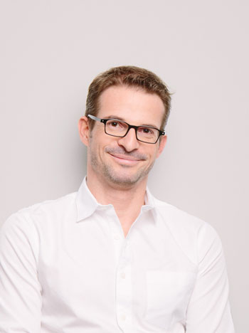
Radiology Center - Dr. Johannes Sailer

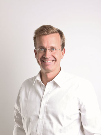
Doz. Dr. Johannes Sailer

Specialist for Radiology

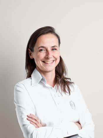
Radiology Center - Dr. Martha Hoffmann

Doz. Dr. Martha Hoffmann

Head of Nuclear Medicine

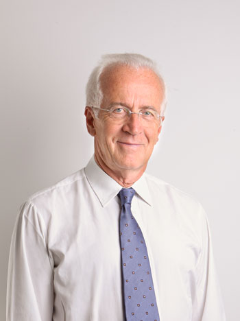
Radiology Center - Dr. Peter Peloschek

Dr. Peter Peloschek

Specialist in radiology

The clinic

Medical devices

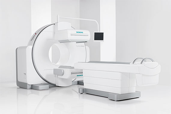
Szintigraphiegerät SPECT Symbia Intevo TM
Scintigraphy is one of the nuclear medicine examinations for which a radioactive substance is administered and a subsequent scan is used to measure the accumulation of this material in certain organs and tissues of the body. The stored amount of the radionuclide provides information about the metabolism of these organs or their function. This method is suitable for localizing inflammation sites, e.g. in the skeleton, but also for measuring and visualizing cardiac muscle perfusion, kidney function and brain metabolism. The Symbia Intevo is designed to perform examinations with the smallest possible amount of radioactive material. The CT scan, which is performed at the same time if necessary, provides additional information that ensures precise and reliable localization of conspicuous areas in the body. The Symbia Intevo is designed in such a way that the examinations can be carried out with the smallest possible amount of radioactive material with extremely fast measurements, yet still enabling an enormously high level of tissue differentiation. The CT scan, which is performed simultaneously if necessary, provides additional information that ensures precise and reliable localization of conspicuous areas in the body. This diagnostic information allows reliable interpretation by the physician and provides the basis for rapid therapy decisions.
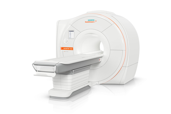
MRI uses a very strong magnetic field and high-frequency radio waves, i.e. without X-rays, to generate high-resolution image data from almost all areas of the body.
Magnetresonanztomographiegerät Magnetom Altea 1,5 T
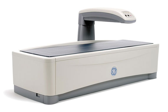
Dual energy X-ray absorptiometry (DXA, formerly also DEXA) is the established reference method for the diagnosis of osteoporosis. Two images are taken with different X-ray energy and subtracted from each other, thus the amount of bone calcium can be detected. Bone density is measured in two areas as standard: Hip joint and lumbar spine and takes about 15 minutes. During this time, you can breathe normally and calmly. Radiation exposure is minimal.
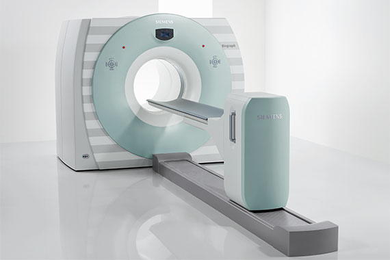
Positron emission tomography (PET) is a modern nuclear medicine procedure that is becoming increasingly important, especially in tumor diagnostics. In combination with a computer tomograph (PET-CT), biochemical and physiological processes in body tissues can be visualized after an injection of weakly radioactively labeled substances. The simultaneously performed CT provides additional information that ensures precise and reliable localization of conspicuous areas in the body. The innovative design of the Biograph 16 TP True V makes it possible to image large regions of the body three-dimensionally and in a very short time in a single session. Due to an additional fourth detector ring (extended axial field of view), the sensitivity of this PET/CT is about 75% better compared to all other systems available on the market, i.e. the examinations can be performed much more gently (less radiation, faster). The innovative design of the Biograph 16 TP True V makes it possible to image large regions of the body three-dimensionally and in a very short time in a single session. This has the great advantage that only small amounts of radioactively labeled substances are required for this purpose without impairing the image quality. Low radiation and short times are of great importance for the patient, especially in the case of frequent control examinations. Due to an additional fourth detector ring (extended axial field of view), the sensitivity of this PET/CT is about 75% better compared to all other systems available on the market, i.e. examinations can be performed much more gently (less radiation, faster).
Positronen-Emissionstomographiesgerät Biograph 16TP
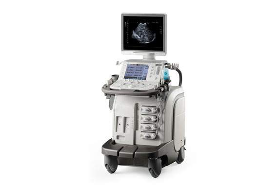
Only ultrasound waves are used in sonography to generate moving images. With the help of the high-resolution transducer and heavy-wave elastography, pathological changes (inflammations, cancers, thyroid nodules) can be detected and assigned with high accuracy.
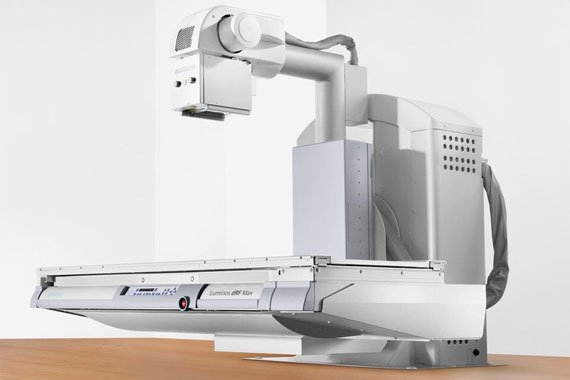
Accurate digital images of internal organs, joints and bones can be made with a small amount of X-ray radiation. If required, short X-ray films (fluoroscopy) can also be taken, for example to examine the mobility of the stomach or intestines.
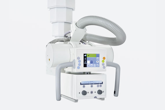
A digital X-ray unit that uses a small amount of X-rays to take high-resolution digital images, especially of joints, for diagnosis and treatment planning in orthopedics and trauma surgery. This X-ray unit provides exceptional imaging flexibility in a wide range of settings with patients standing, sitting and lying down.
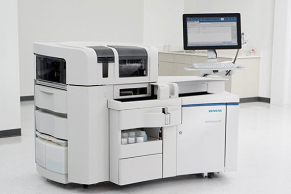
Our in-house thyroid laboratory uses a Siemens Advia Centaur immunoassay analyzer to determine your thyroid hormones (TSH, T4, etc.) immediately on-site with the highest possible accuracy.

History

Foundation of the X-ray ordination as "X-ray department in the sanatorium of Dr. Anton Löw as evidenced by two publications by Guido Holzknecht from 1902/03. Guido Holzknecht is considered the founder of the "Viennese School of Radiology". Until World War II, the bearers of such famous names as the already mentioned Guido Holzknecht, Ludwig v. Schwarda or Julian Bartsch worked here. After the reconstruction of the Vienna Private Clinic after the end of the war, the X-ray ordination was reopened by Josef Palugyay. His successor was then Ludwig Psenner.

Prim. Univ. Prof. Dr. Heinrich Czembirek took over the ordination of Prof. Ludwig Psenner (1909 - 1986).

OA Dr. Med. Univ. Elisabeth Kalinowski led the X-ray coordination at the Vienna Private Clinic together with Prof. Czembirek.

Dr. Philipp Peloschek has been in charge of magnetic resonance imaging at the X-ray department of the Vienna Private Clinic.

OA Dr. Med. Univ. Elisabeth Kalinowski led the X-ray coordination at the Vienna Private Clinic together with Privatdozent Dr. Philipp Peloschek. In this year, private lecturer Dr. Philipp Peloschek began planning and developing the Radiology Center.

The opening of the Radiology Center took place, and thus for the first time in Austria a PET/CT was put into operation outside of a focal hospital.

Prof. Dr. Martha Hoffmann took over the management of the PET/CT and all other nuclear medicine tasks.

PD Dr. Johannes Sailer joined the Radiology Center team as a partner.

We take over the complete diagnostic management from symptom to needle biopsy with our Care Center if required: Thyroid Care Center, Breast Care Center, Heart Care Center, Prostate Care Center.

FAQs

The Radiology Center is located directly opposite the renowned “Goldenes Kreuz” maternity clinic and women’s health center in Vienna’s 9th district and is connected underground to the renowned “Wiener Privatklinik”. However, it is particularly important to emphasize that it is an independent enterprise and has been frequented by many thousands of outpatients from all over Vienna, Lower Austria, Burgenland and all over the world for decades.> Directions

Patients expect one thing above all from the wide range of imaging procedures available to medicine today: more security for their health. The Radiology Center offers state of the art technology, which was previously limited to hospitals, combined with the benefits of an optimally run specialist practice, detailed discussions with patients, individual care and extremely short waiting times. In the Radiology Center, the focus is on the patient: A very important principle for the medical, technical and organizational offer at the Radiology Center is orientation towards the patient and their individual needs. Additionally, the most modern radiology and nuclear medicine awaits you all under one roof. > About us

Appointment scheduling Monday to Thursday from 9:00 AM to 5:00 PM and Fridays from 9:00 AM to 3:00 PM.
We are happy to answer your email inquiries at office@radiology-center.com

If required, it is also possible to arrange appointments for an MRI (Magnetic Resonance Imaging) on Monday, Tuesday, and Thursday between 5:00 PM and 7:00 PM.

Many of the examinations at the Radiology Center can be settled with the Austrian social insurance (health insurance/e-card).

When offsetting our doctor’s office of choice for X-rays and ultrasound with health insurance (reimbursement of costs by social insurance of 80%), our secretariat will, if desired, take over the submission of the reimbursement of costs.

Since small insurance companies charge a deductible of 20%, additional costs only arise for WGKK patients (approx. EUR 3 to 19 depending on the examination). In addition to the health insurance contract services, there are many examinations that are not or only partially paid for by health insurance.

We will inform you in good time about any costs incurred and support you in processing reimbursements from the health insurance company. Private services can be paid for on site in cash, with a debit or credit card.

You need a referral from your doctor for an appointment at the Radiology Center for all examinations that use ionizing radiation or a contrast medium (X-ray, computed tomography).

No, in the radiology center you will receive your appointment in a few days time.

Do you have any questions and haven’t reached us?
Just write us an email or leave us a message and/or your telephone number.
Our employees will contact you immediately.

office@radiology-center.com (or click on the contact icons at the top right)

Personal support from our core team is important to us: of course, you can have your radiological examinations personally carried out by private lecturer Dr. Johannes Sailer or private lecturer Dr. Philipp Peloschek or Prim.i.R. dr Peter Peloschek to carry out. The nuclear medical examinations are carried out by Prof. Hoffmann.

A group of highly qualified experts is often required to offer all radiological options at the highest level. We pride ourselves on being able to draw on the expertise of a group of highly qualified experts when needed.
> Our Team