Scintigraphy

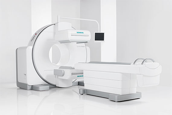
Scintigraphy is the measurement and imaging of the radioactively labeled substances (radiopharmaceuticals) described above in the body using a gamma camera to visualize organ function. This takes the form of individual images (e.g., thyroid scintigraphy), whole-body images (e.g., skeletal scintigraphy), or serial (dynamic) images (e.g., renal scintigraphy). Scintigraphy can be used to examine the metabolic function of virtually all organ systems.

Depending on the organ to be examined, scintigraphy (or SPECT) requires a different accumulation time: in bone scintigraphy, it takes about two to three hours for the radioactive substance to accumulate predominantly in the skeletal system; in thyroid scintigraphy, it takes only about 20 minutes. For some examinations, such as checking for side-separated renal function, the scintigraphy begins immediately after the tracer is administered.

Certain examinations require special preparation, about which patients are informed in detail by the referring physician or when making the appointment. For example, cardiac scintigraphy, which is used to diagnose coronary heart disease (CHD, precursor of a heart attack), requires (pharmacological) stress on the heart. This can be used to determine whether blood flow to the heart muscle is reduced in a certain area under stress conditions (stress, physical exertion). In cases of already known and successfully treated CHD, this examination method is a reliable (non-invasive) option for follow-up.

The nuclear medicine examinations are performed by Prof. Hoffmann.

Scintigraphy is one of the nuclear medicine examinations for which a radioactive substance is administered and a subsequent scan is used to measure the accumulation of this material in certain organs and tissues of the body. The stored amount of the radionuclide provides information about the metabolism of these organs or their function. This method is suitable for localizing inflammation sites, e.g. in the skeleton, but also for measuring and visualizing cardiac muscle perfusion, kidney function and brain metabolism. The Symbia Intevo is designed to perform examinations with the smallest possible amount of radioactive material. The CT scan, which is performed at the same time if necessary, provides additional information that ensures precise and reliable localization of conspicuous areas in the body. The Symbia Intevo is designed in such a way that the examinations can be carried out with the smallest possible amount of radioactive material with extremely fast measurements, yet still enabling an enormously high level of tissue differentiation. The CT scan, which is performed simultaneously if necessary, provides additional information that ensures precise and reliable localization of conspicuous areas in the body. This diagnostic information allows reliable interpretation by the physician and provides the basis for rapid therapy decisions.
Szintigraphiegerät SPECT Symbia Intevo TM

Scintigraphy is the measurement and visual representation of the radioactively labeled substances (radiopharmaceuticals) described above in the body, using a gamma camera to depict organ function. This is done in the form of individual images (e.g., in thyroid scintigraphy), whole-body images (e.g., in skeletal scintigraphy), or serial (dynamic) images (e.g., in renal scintigraphy). With the help of scintigraphy, practically all organ systems can be tested for their metabolic function.

Depending on the organ to be examined, scintigraphy (or SPECT) requires a different accumulation time: in bone scintigraphy, it takes about two to three hours for the radioactive substance to accumulate predominantly in the skeletal system; in thyroid scintigraphy, it takes only about 20 minutes. For some examinations, such as checking for side-separated kidney function, the scintigraphy begins immediately after the tracer is administered.

Many of the examinations at the Radiology Center can be settled with the Austrian social insurance (health insurance/e-card).

When offsetting our doctor’s office of choice for X-rays and ultrasound with health insurance (reimbursement of costs by social insurance of 80%), our secretariat will, if desired, take over the submission of the reimbursement of costs.

Since small insurance companies charge a deductible of 20%, additional costs only arise for WGKK patients (approx. EUR 3 to 19 depending on the examination). In addition to the health insurance contract services, there are many examinations that are not or only partially paid for by health insurance.

We will inform you in good time about any costs incurred and support you in processing reimbursements from the health insurance company. Private services can be paid for on site in cash, with a debit or credit card.

Certain examinations require special preparation, about which patients are informed in detail by the referring physician or when making the appointment.

Example: If you are coming for a cardiac scintigraphy, which serves to clarify coronary heart disease (CHD, precursor of a heart attack), you must be fasting for 6 hours and you must not have had any caffeine 12 hours before the examination (water and medication are, however, permitted).

Salivary gland scintigraphy is used to assess the function of the major salivary glands of the head (parotid gland, submandibular gland). In cases of suspected Mb. Sjögren’s disease, salivary gland scintigraphy may be useful to confirm the diagnosis and to assess the severity of the disease. Furthermore, salivary gland scintigraphy is useful in the clarification of xerostomia, as well as before and after radiotherapy (external neck irradiation).

Tc99m sestamibi parathyroid scintigraphy is used in cases of known primum or, in exceptional cases, sec. Hyperparathyroidism for preoperative localization diagnosis. This allows minimally invasive surgery.